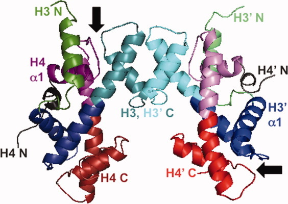
Figure 1.

3D domain swapping and the structure of histone oligomers. Ribbon diagram of the (H3-H4)2 tetramer derived from the structure of the nucleosome core particle (1AOI.pdb).2 The H3-H3′ four-helix bundle tetramer interface is at the top of the figure. Some regions of the poorly structured N-terminal tails have been omitted for clarity. The H3 monomers are shown in green (N-terminal tail and helix) and blue to cyan (histone fold); the H4 monomers are shown in gray (N-terminal tail and helix) and purple to red (histone fold). The two of the four β-bridges are indicated by arrows. The protein structure was rendered with the program Mac-PyMol (DeLano Scientific LLC, Portland, OR).
