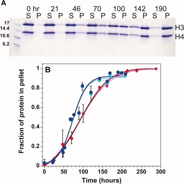
Figure 2.

Precipitation gel assay for histone fibrillation. A: Coomassie Blue stained SDS-polyacrylamide gel monitoring the time-dependence of precipitation of H3 + H4 monomers initially unfolded in 10 mM HCl. S, supernatant, P, pellet. B: Densitometer quantitation of the fraction of protein in the pellet for H3 (lighter symbols, dotted line) and H4 (darker symbols, solid line) starting from unfolded H3 + H4 (red diamonds) or from folded tetramer at pH 7.2 (blue circles). The lines represent fits of the data to Eq. (1). Error bars at one standard deviation from multiple experiments are shown or are smaller than the size of the data points. Final conditions: 1M NaCl, 50mM acetic acid/sodium acetate, and 50 mM phosphoric acid/NaH2PO4, pH 5, 23°C and 30 μM each of H3 and H4 monomers. [Color figure can be viewed in the online issue, which is available at wileyonlinelibrary.com.]
