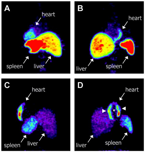
Figure 6. Myocardial homing and biodistribution of 18F-FDG–labeled BMCs.
Left posterior oblique (A) and left anterior oblique (B) views of chest and upper abdomen of patient 2 taken 65 minutes after transfer of 18F-FDG–labeled, unselected BMCs into left circumflex coronary artery. BMC homing is detectable in the lateral wall of the heart (infarct center and border zone), liver, and spleen. Left posterior oblique (C) and left anterior oblique (D) views of chest and upper abdomen of patient 7 taken 70 minutes after transfer of 18F-FDG–labeled, CD34-enriched BMCs into left anterior descending coronary artery. Homing of CD34enriched cells is detectable in the anteroseptal wall of the heart, liver, and spleen. CD34+ cell homing is most prominent in infarct border zone (arrowheads) but not infarct center (asterisk) (with permission from Circulation – ref. 89).

