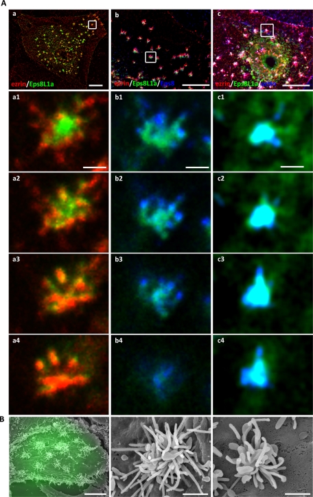
FIGURE 4:
Coexpression of ezrin and Eps8L1a induces microvillar clustering at the apical surface of LLC-PK1 cells. LLC-PK1 cells were cotransfected with plasmids coding for ezrin-VSVG and GFP-Eps8L1a. (A) Immunofluorescence was performed with an anti-VSVG antibody to detect ezrin (a, b, c, a1–a4; red) and an anti-Eps8 antibody (b, b1–b4; blue). Actin was detected with phalloidin (c, c1–c4; blue). Images correspond to a maximum-intensity Z-projection performed after deconvolution. Bottom, four successive slices of the clusters in the insets from a–c are shown from the bottom (a1, b1, c1) to the top of the structure (a4, b4, c4). Eps8L1a is mainly localized in the enlarged membrane structures (see scanning electron microscopy) and ezrin in the microvilli that originate from them. Bars, a–c, 10 μm; a1–c4, 1 μm. (B) CL-SEM. Low (left) and high (middle and right) magnification of the surface of LLC-PK1 cells expressing ezrin-VSVG and GFP-Eps8L1a. Clusters from two different cells are shown at high magnification. Transfected cells are monitored by fluorescence through GFP fluorescence. Bars, left, 10 μm; middle and right, 1 μm.

