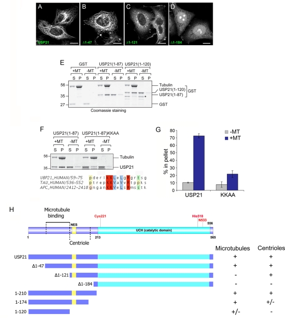
FIGURE 5:
Mapping the requirements for USP21 localization and binding to microtubules. (A–D) HeLa cells were transfected for 24 h with full-length GFP-USP21 wild type or the indicated N-terminal truncation mutant. Scale bar, 10 μm. Note that this localization was also confirmed in U2OS cells. (E) In vitro spin-down assay for direct binding to purified microtubules (MTs). Binding of both USP21 N-terminal fragments (1–87 and 1–120) to microtubules is evidenced by redistribution from supernatant (S) to pellet fraction (P). Asterisk indicates a shorter truncation, which has lost microtubule binding. (F) Alignment of USP21 residues 59–75 with motifs in tau and APC proteins: mutation of matching lysine residues diminishes in vitro binding to microtubules. (G) Quantitation of data presented in F. Bars indicate range of two distinct experiments. (H) Summary of constructs used and properties reported in this figure and Supplemental Figures S4 and S5.

