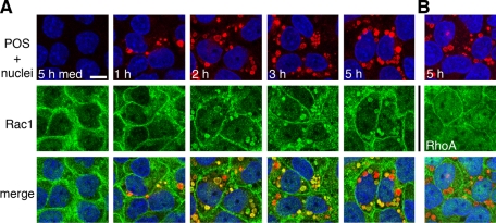
FIGURE 2:
Rac1 redistributes to colocalize with phagocytosed POS. RPE-J cells on glass coverslips were challenged with medium for 5 h (5 h med) or with Texas red–stained POS for different lengths of time (as indicated) before immunolabeling of Rac1 (A) or RhoA (B). Single x-y confocal scans show: top, overlay of POS (red) with RPE nuclei (blue); middle, GTPases (green); or bottom, overlay of POS, nuclei, and GTPase. Scale bar (for all fields): 10 μm.

