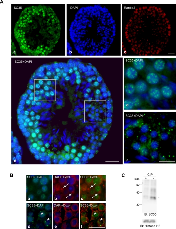
FIGURE 1:
Distribution of phosphorylated SR proteins in adult mouse testis. (A) The distribution of phosphorylated SR proteins and expression of Ranbp2 are developmentally regulated. Representative views of cross-sections of seminiferous tubules stained with the mouse monoclonal antibody SC35 (a), DAPI (b), and anti-Ranbp2 (c). (d–f) Merged images of SC35 and DAPI. (e, f) Enlarged pictures of the boxed areas in d. SC35 antigens were present in a speckled pattern in the nucleus of spermatocytes (e), whereas they formed granular structures in the cytoplasm in the spermatids (f). The differentiation stage was determined based on morphology under DAPI staining. (B) An adult mouse testis section was immunostained with SC35 (green) and antibodies against Ddx4 for nuage/germinal granules (red). Nuage/germinal granules (arrows) were not recognized by SC35 (a–c). In addition, cells with SC35-stained granules (arrowheads) did not form nuage/germinal granules (d–f). Bars, 20 μm (A, a–d), 10 μm (A, e–f, and B). (C) Immunoblot analysis of cell lysate prepared from adult mouse testis. Antibody SC35 recognizes mouse srsf2 (asterisk) in this tissue, but it fails when lysate is treated with phosphatase. Histone H3 serves as a loading control.

