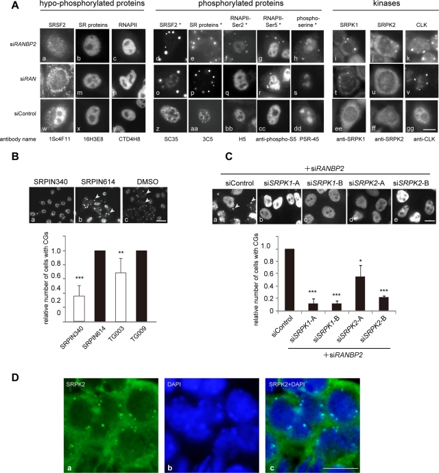
FIGURE 5:
Phosphorylation of SR proteins is involved in CG generation. (A) CG localization of proteins correlated with their phosphorylation status. Cells were stained with antibodies recognizing hypophosphorylated forms (left) and the corresponding phosphorylated forms (middle) of the indicated proteins. Phosphoepitopes are denoted with an asterisk (Thompson et al., 1989; Bregman et al., 1995; Cavaloc et al., 1999). Cells were also stained with antibodies against SR protein kinases (right). (B) RANBP2-knockdown cells were treated with SR protein kinase inhibitors (SRPIN340 and TG003; unfilled bars) or control compounds (SRPIN614 or TG009; filled bars; Kuroyanagi et al., 1998; Muraki et al., 2004) for 20 h and immunostained with SC35. Arrowheads indicate CGs (top). Relative numbers of RANBP2-negative CG-containing cells are shown on the graph. Inhibition of SRPKs led to a reduction in CG generation. The values are the means and SDs of three independent experiments of >250 cells each. **p < 0.01, ***p < 0.001 vs. control compounds. (C) Cells were treated with the indicated combinations of siRNAs and were immunostained with the monoclonal antibody mAb104. Relative numbers of RANBP2-SRPK–negative, CG-containing cells are shown. For knockdown of SRPK1 and SRPK2, two siRNAs with different target sequences were used. Depletion of SRPKs significantly reduced CG generation in RANBP2-knockdown cells. The values are the means and SDs of three independent experiments of >100 cells each. *p< 0.05, ***p < 0.001 vs. controls. (D) Srpk2 was also found in cytoplasmic granular structures in mouse seminiferous tubules. Bars, 10 μm (A–C), 5 μm (D).

