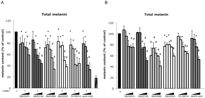
Figure 1. Effect of pyridinyl imidazoles compounds on melanin synthesis in B16-F0 melanoma cells.
(A) Following incubation with α-MSH (0.1 µM) and increasing concentrations (1, 2.5, 5, 10, 20 µM) of pyridinyl imidazoles for 72 h, the extracellular and intracellular levels of melanin were determined separately by measuring the absorbance at 405 nm. Standard curves of synthetic melanin were used to extrapolate the absolute values of melanin content. The total amount of melanin was calculated for each experimental point by adding the extracellular and intracellular melanin values after normalization for protein content. Total melanin produced at the end-point by control (DMSO-treated cells) and hormone-stimulated cells (α-MSH plus DMSO-treated cells) is reported for comparison. (B) B16-F0 cells were also treated with pyridinyl imidazoles compounds for 96 h in absence of α-MSH. Results are expressed as percentage of untreated control samples. The data show the mean±SD of three experiments performed in duplicate. *P≤0.05; #P≤0.01 versus control.

