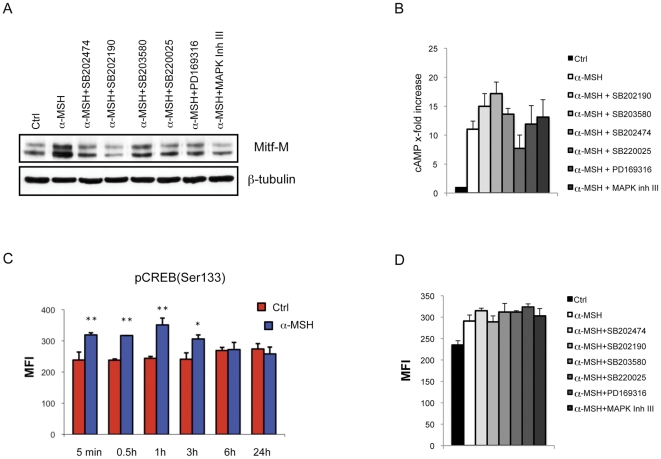
Figure 3. The effect of pyridinyl imidazoles on cAMP/PKA/CREB signal transduction.
(A) Expression of Mitf in B16-F0 cells after 6 h of treatment with α-MSH (0.1 µM) in presence or not of PI compounds (SB202474, SB202190, SB203580, SB220025, PD169316 20 µM: MAPK Inh III 10 µM). Total cellular proteins (30 µg/lane) were subject to 10% SDS-PAGE. Variation of loading was determined by blotting with anti-β-tubulin antibody. Western blot assays are representative of at least three experiments. (B) Concentration of cAMP of control and treated cells were determined using the cAMP bioluminescent assay. Following incubation with α-MSH (0.1 µM) in presence or not of PI compounds, the cAMP levels were measured and compared to the untreated control samples. The results are the mean±SD of three experiments performed in duplicates. (C) Analysis of the time-dependent effect α-MSH treatment on CREB level of phosphorylation. Cells were stained with anti-phospho-CREB-PE (Ser133), and then analyzed measuring median fluorescence intensity (MFI) in duplicates. Histogram represents means ± SD of MFI of three independent experimets. (D) Comparative analysis of CREB-Ser133 level of phosphorylation in untreated cells, α-MSH-treated cells and α-MSH plus PI coumpond-treated cells.

