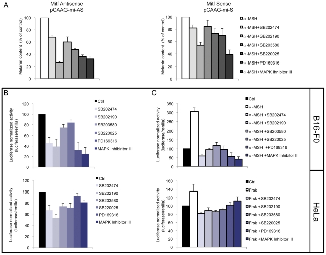
Figure 5. Analysis of the role of Mitf expression in pyridinyl imidazoles-dependent melanogenesis inhibition.
(A) To B16-F0 melanoma cells were transiently transfected with a plasmid encoding for Mitf cDNA (pCAAG-mi-S) or a control construct carrying Mitf cDNA in antisense orientation (pCAAG-mi-AS). Following incubation with α-MSH (0.1 µM) in presence of pyridinyl imidazoles (SB202474, SB202190, SB203580, SB220025, PD169316 20 µM: MAPK Inh III 10 µM) for 72 h, or not, the extracellular and intracellular levels of melanin were determined as described above. The data show the mean±SD of three experiments performed in duplicate. (B–C) Analysis of luciferase activity of the Mitf melanocyte-specific promoter (M promoter) in the presence of pyridinyl imidazoles in B16-F0 and HeLa cells. Twenty-four hours after transient transfection cells were treated with PI compounds in presence (C) or not (B) of α-MSH (0.1 µM) (B16-F0) or forskolin (1 µM) (HeLa). Luciferase activity was assayed after 6 h of treatment. Firefly luciferase activity, normalized to the corrisponding renilla luciferase activity was expressed as fold change compared with control cells. Values represent mean ± SD of three representative experiments performed in duplicate.

