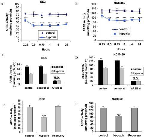
Figure 1. ARSB activity reduced by hypoxia in BEC and NCM460 cells.
A. ARSB activity in the bronchial epithelial cells (BEC) was measured following exposure to 10% oxygen environment for 0.25, 0.5, 0.75, 1, 4, and 24 hours. ARSB declined significantly in activity by 0.25 hours (p<0.001), and remained at this level for 24 hours, in contrast to control cells under normoxic conditions. B. Similarly, following exposure to 10% O2, ARSB activity declined in the NCM460 cells and was significantly reduced at 0.25 h (p<0.05). ARSB declined further by 30 minutes (p<0.01), reaching maximum reduction by 0.75 h (p<0.001), and remained at this level for 24 h, in contrast to control cells. C. The combination of hypoxia (10% O2×4 h) and ARSB silencing in the BEC produced no further decline than that achieved by ARSB silencing alone. D. Similarly, in the NCM460 cells, the combination of ARSB silencing and hypoxia (10% O2×4 h) did not lead to further decline in the ARSB activity than that produced by ARSB siRNA alone. E. In the BEC, return to normoxia for 4 h after hypoxia (10% O2×4 h) restored the baseline ARSB activity. F. In the NCM460 cells, return to normoxia for 4 h after 4 h of 10% O2 restored the baseline ARSB activity. [ARSB = arylsulfatase B; BEC = bronchial epithelial cell; N.D. = no difference].

