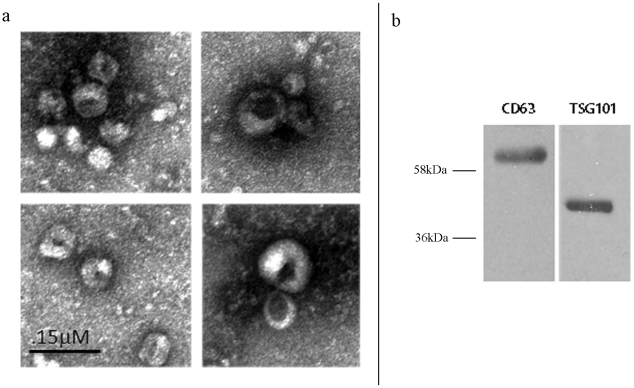
Figure 1. Confirmation that the ultracentrifugation pellet contains exosomes.
a Electron microscopy of the ultracentrifugation pellet from serum shows the characteristic spherical shape and size (50–100 nm) of exosomes b. Western blot shows strong staining of the ultracentrifugation pellet with the exosomal membrane markers anti cd63 and TSG101.

