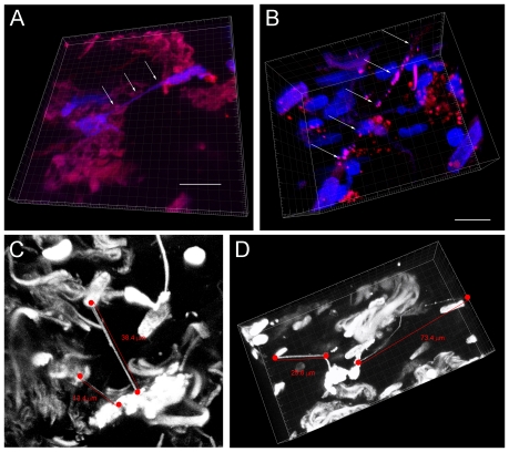
Figure 6. Tunneling nanotubes are present in solid tumors resected from patients with malignant pleural mesothelioma and lung adenocarcinoma.
Confocal microscopy was performed and 3-dimensional images constructed using Imaris Viewer. a) Two cells from a mesothelioma solid tumor are connected by a nanotube in three-dimensional plane. b) A long TnT is noted in another mesothelioma tumor specimen. c) Lung adenocarcinoma tumor specimen also manifesting multiple intact nanotubes of various lengths. d) Nanotubes present in a lung adenocarcinoma tumor sample vary in length, width, and extent of curvature. Multiple “blebs” are noted in the lengthy, curved nanotube on the right, measuring at least >70 µm. Scale bars: a) 15 µm, b) 15 µm; c) and d) as noted in the figure.

