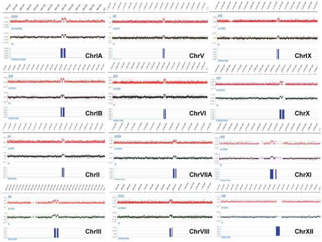
Figure 4. TgChromo1 binds to peri-centromeric heterochromatin.
ChIP on chip was performed with the TgChromo1 antibody (anti-CHD1, red) or the anti-HA antibody (HA, black) and hybridized on a genome-wide tiling microarray. The regions of enrichment for H3K9me3 [12] are represented in blue. A snapshot of the 12 chromosomes where a centromere was identified [12] is presented. ChIP on chip signals are represented as a log2 ratio of the signal given by the immunoprecipitated DNA over the input and plotted according to the genomic position of the oligonucleotide.

