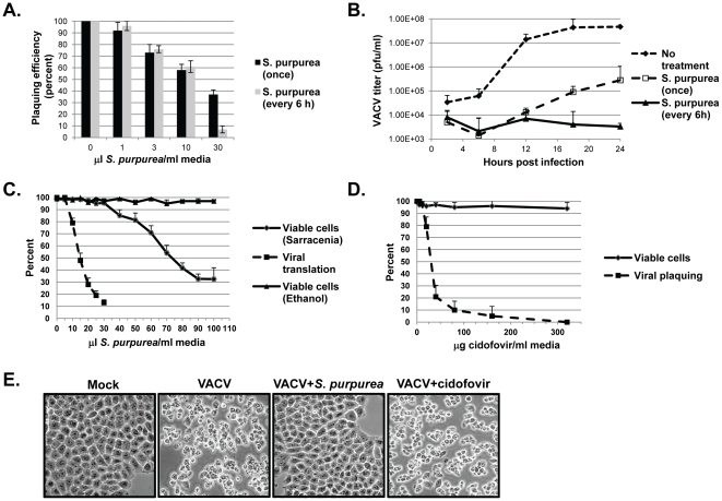
Figure 1. The effect of S. purpurea extracts on VACV replication.
A) RK-13 cells were infected with 150 pfu of VACV followed by the addition of the indicated concentration of S. purpurea extract to the cell culture media. Cells were treated one-time only with the extract (black bars) or every 6 hours with fresh extract (gray bars). After 48 hours, plaques were visualized and quantified. Error bars represent standard deviation (n = 3). B) RK-13 cells were infected with VACV at an multiplicity of infection (MOI) = 10 followed by addition of ethanol/glycerol carrier (closed diamonds) or the addition of 25 microL S.purpurea extract/ml media (open squares and closed triangles). Cells were treated one-time only with the extract (open squares) or every 6 hours with fresh extract (closed diamonds). Cells were harvested at the indicated times and viral titers determined. Error bars represent deviation between assays (n = 2). C) For viral translation levels (closed squares), HeLa cells were infected with VACV at an MOI = 10 followed by the addition of the indicated concentrations of S. purpurea extract/ml media. At 6 HPI, cell lysates were prepared, the VACV E3L protein detected by Western blot, and quantified. For cell viability, HeLa cells were treated with the indicated concentrations of S. purpurea extract (closed diamonds) or ethanol/glycerol carrier (closed triangles) for 6 hours and the number of viable cells determined by a trypan blue exclusion assay. D) For viral plaque formation (closed squares), HeLa cells were infected with VACV (approx. 200 pfu) followed by the addition of the indicated concentrations of cidofovir to the media. After 48 hours, plaques were visualized and quantified. Error bars represent standard deviation (n = 3). For cell viability, HeLa cells were treated with the indicated concentrations of cidofovir (closed diamonds) for 48 hours and the number of viable cells determined by a trypan blue exclusion assay. E) HeLa cells were mock-infected or infected with VACV at an MOI = 10 followed by the immediate addition of media, 25 microL S. purpurea extract/ml media, or 320 microg cidofovir/ml media. At 4 HPI, the cell monolayers were photographed.

