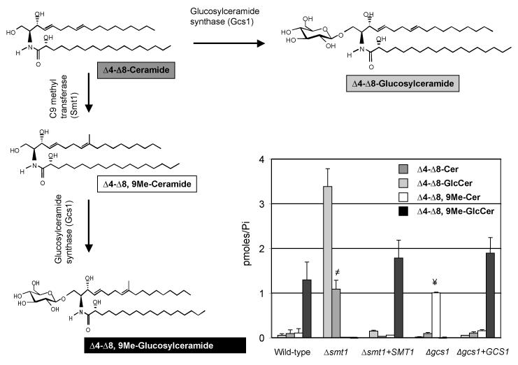
Figure 2. Deletion of Smt1 produces a strain that make only un-methylated GlcCer.
Analysis of specific lipids extracts of WT, Δsmt1, Δsmt1+SMT1, Δgcs1, and Δgcs1+GCS1 cells along with the structure illustration of α-OH-Δ4-Δ8-ceramide, α-OH-Δ4-Δ8,9-methyl-ceramide, α-OH-Δ4-Δ8,9-methyl-GlcCer and α-OH-Δ4-Δ8-GlcCer. The WT along with Δsmt1+SMT1 reveals almost identical amounts of α-OH-Δ4-Δ8,9-methy-GlcCer but negligible amounts of α-OH-Δ4-Δ8-ceramide and its methylated product α-OH-Δ4-Δ8,9-methyl-ceramide. However, the Δsmt1 does not produce methylated GlcCer and accumulates α-OH-Δ4-Δ8-ceramide. In contrast, Δgcs1 accumulated α-OH-Δ4-Δ8,9-methyl-ceramide and lacked methylated and unmethylated GlcCer. All results were means ± SD of three independent experiments. ≠P<0.05, Δsmt1 versus wild-type; ¥P<0.05, Δgcs1 versus WT.

