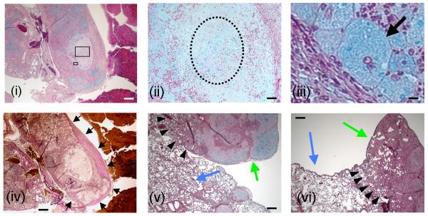
Figure 5. Histopathological analysis of Δsmt1-infected lungs.
Lung sections of Δsmt1-infected mice were performed at 90 days of infection. They reveal distinct formation of granulomas within which Δsmt1 cells are contained. Panels i, ii, iii, v and vi are Movat stained sections, whereas panel iv is stained with VVG and depicts the same area illustrated in i. Boxed areas in 5i are magnified in 5ii and 5iii. C. neoformans Δsmt1 cells are stained blue in i, ii, iii, v and vi) are mostly contained within giant macrophage(s) (arrow in iii) or within a necrotic zone (dotted circle in ii). The granulomatous response is surrounded by collagen fibers stained pink-to-dark red (black arrows in iv). Normal lung tissues are indicated by blue arrows in v and vi, in close proximity of granuloma (green arrows in v and vi). Scale bars: 500 μm (i, iv, v and vi); 50 μm (ii); 10 μm (iii).

