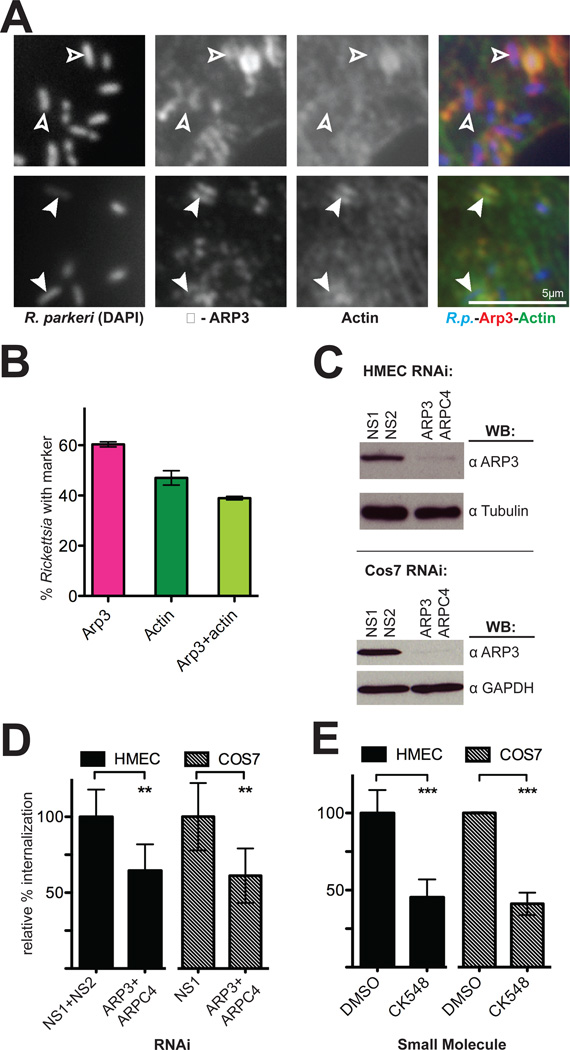
Figure 5. The Arp2/3 complex is recruited to invading R. parkeri and is required for efficient invasion.
(A) Bacteria (left, stained with DAPI, blue in merge) are surrounded by Arp3 (middle, anti-Arp3 immunofluorescence, red in merge) and actin (right, anti-RFP immunofluorescence of Lifeact-mCherry, green in merge). Open arrowheads indicate R. parkeri associated with Arp3 protein only, filled arrowheads indicate R. parkeri surrounded by both actin and Arp3. (B) Percentage of Rickettsia associated with Arp3 and actin. Pink, mean association with Arp3; dark green, mean association with actin; light green, subset of cells associated with both Arp3 and actin. For (A–B), HMEC-1 cells were transfected with pLifeact-mCherry for 48 h before infection and fixed 10 min post-infection. (C) Western blots of HMEC-1 and COS-7 cell lysates at 48 h post-transfection with the indicated siRNAs, corresponding to one experiment represented in (D). (D) Relative percent internalization at 15 min post-infection with R. parkeri in either HMEC-1 (solid bars) or COS-7 (hashed bars) cells. (E) Relative percent internalization at 15 min post-infection with R. parkeri within HMEC-1 or COS-7 cells treated for 30 min prior to infection with either 1% DMSO or 100 µM CK548. (D) and (E) are the mean of at least three independent experiments and were normalized with non-specific RNA-transfected cells (D) or DMSO-treated cells (E) set as 100%. Abbreviations: NS1, nonspecific control RNA 1; NS2, nonspecific control RNA 2; ** p<0.01, *** p<0.001, versus indicated control values by unpaired Student’s t-test.

