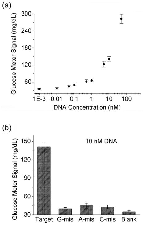
Figure 4.
(a) Detection of a 35-mer HBV DNA fragment using a PGM (the signal plot of a DNA-free sample cannot be shown in the logarithmic chart, which is almost the same as that of the sample with 1 pM target DNA). (b) Single mismatch selectivity of the method. The detection was conducted in 0.15 M sodium phospate buffer, pH 7.3, 0.25 M NaCl, 0.05% Tween-20.

