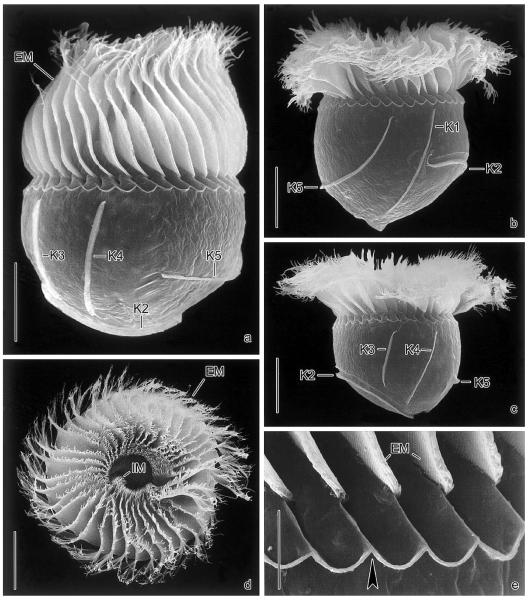
Fig. 2.
Pelagostrobilidium neptuni in the scanning electron microscope. (a–c) Lateral views during ~360° rotation about main cell axis, beginning with the dorsal aspect. (d) Top view showing the conspicuous oral apparatus. (e) Intermembranellar ridges. Arrowhead marks crenated border. EM, external membranelles; IM, internal membranelles; K1–5, somatic kineties. Scale bars 20 μm (a–d) and 5 μm (e).

