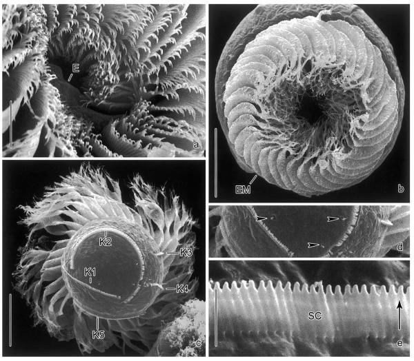
Fig. 3.
Pelagostrobilidium neptuni in the scanning electron microscope. (a) Detail of adoral zone of membranelles and peristomial field. Note the endoral. (b) Top view. In motionless cells, the external membranelles are arranged like an iris diaphragm. (c, d) Posterior polar view and detail of cell surface. The cortical granules are probably extrusive, as they occasionally penetrate the cell surface (arrowheads). (e) Detail of somatic kinety. The ~2 μm long cilia are directed to the left (arrow) due to the kinetal lips covering their proximal portion. E, endoral; EM, external membranelles; K1–5, somatic kineties; SC, somatic cilia. Scale bars 20 μm (b, c), 10 μm (a), and 2 μm (e).

