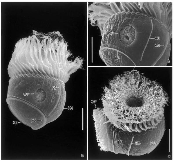
Fig. 5.
Pelagostrobilidium neptuni, dividers in the scanning electron microscope. (a, b) Middle dividers showing the opisthe's oral apparatus in a subsurface pouch on the left cell side anterior to kinety 2. The daughter's external membranelles are arranged like an iris diaphragm. (c) Oblique top view of a late divider showing the evaginated new adoral zone of membranelles. The proter's and opisthe's oral apparatus form a right angle, i.e., the opisthe's right-side faces the proter's left. K1–5, somatic kineties; OP, oral primordium. Scale bar 20 μm.

