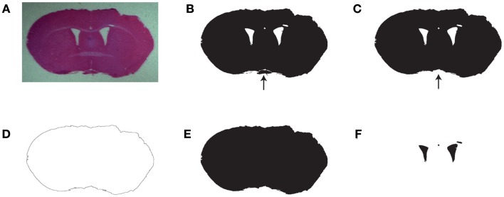
Figure 1.
Work process used in SectionToVolume for the automated identification of tissue and ventricles. (A) Original image from a brain-injured animal stained with H&E. (B) Pixels with tissue color identified. (C) The largest continuous area of tissue pixels. The arrows in (C,D) point to an area of tissue, which was not continuous with the main part of the section. If present, it would have prevented proper detection of the perimeter. (D) The perimeter of the largest object. (E) All pixels enclosed by the perimeter of the section. (F) The ventricles are identified as the pixels which are white in (B) and black in (E). Note that some areas with low staining intensity are assigned as part of the ventricles. This must be manually corrected.

