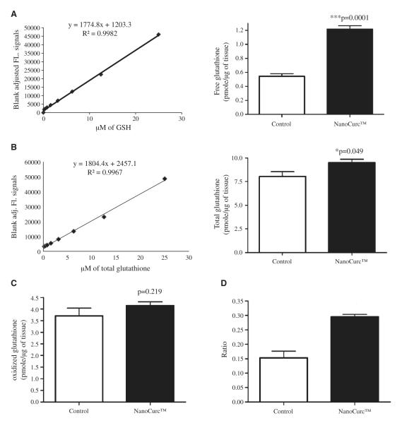
Fig. 5.
(A) A sensitive fluorescence based sensitive assay was used to assess free glutathione levels in the brain lysate of controls and NanoCurc™ treated mice. Fluorescence signals were normalized with the protein content of the brain lysate samples and plotted as pmole/μg. A significant increase in the levels of GSH was observed in the lysate of the treated mice versus controls. A standard curve with known amount of GSH was also shown in the left. (B) The levels of total glutathione (GSH+GSSH) was measured by the same fluorescent based assay as shown in Fig. 5A, which revealed a significant increase in the levels of total glutathione in the lysate of NanoCurc™ treated mice versus controls. (C) Levels of oxidized GSH were measured and no difference in the levels of oxidized GSH was observed in the lysate of NanoCurc™ treated mice versus controls. (D) A significant increase was observed in the ratio of GSH to GSSH in the brain lysate of NanoCurc™ treated mice versus controls, indicating an effective redox cellular environment by NanoCurc™ treatment to athymic mice.

