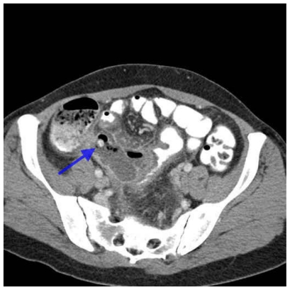
Figure 3.
Computed Tomography
Contrast enhanced axial CT of the abdomen demonstrates dilatation and inflammation of the appendix with surrounding soft tissue stranding. Gas bubbles are seen within and outside the appendiceal lumen, medial to the cecum, consistent with perforated appendicitis. The previously seen 1 cm fecolith is again identified in the proximal appendix (arrow).

