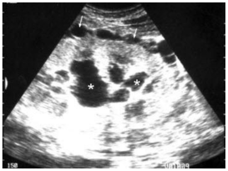
Figure 1.
Abdomen ultrasound of a 22 male patient with renal lymphangiectasia, done by Siemens-sienna machine using a convex transducer of 3.5 MHz. The grayscale coronal view at the right flank shows enlarged right kidney, about 14 cm long, with renal sinus cysts (asterisks) and perinephric collections (arrows).

