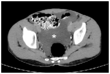
Figure 8.
Follow up abdomen CT scan of a 22 male patient with renal lymphangiectasia. The examination was done by Brilliance 64 Philips machine; kV 120.0, mA 246 and 3 mm slice thickness. Oral contrast and 70 ml IV contrast (Ultravist) were given. The axial contrast enhanced section in the excretory phase at level of the pelvis shows relative improvement in the ascites (asterisk).

