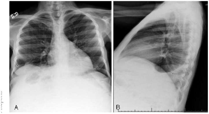
Figure 1.
47 year old woman with a chief complaint of giant abdominal mass and a final diagnosis of uterine leiomyoma. (a) PA Chest X-ray with insufficient inspiration likely due to elevation of the diaphragm. There is also sub-segmental atelectasis. (b) Lateral Chest X-ray showing mild to moderate left sided pleural effusion.

