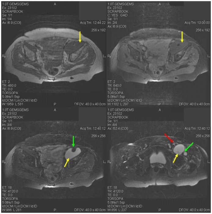
Figure 1.
81 years-old female patient with atypical iliopsoas bursitis. T1 weighted axial image (a), T1 weighted axial contrast enhanced image (20 ml gadolinium was injected) with fat suppression (b) and T2 axial weighted image (c) at the level of the lower pelvis and T2 weighted axial image at the level of the hip joint demonstrating a non-enhancing cystic lesion (yellow arrow) lying in the left iliac fossa coursing at a lower level below the iliopsoas tendon (green arrow). At the level of the hip joint the cystic lesion (yellow arrow) is demonstrated coursing below the iliopsoas tendon and totally encasing it (green arrow). Femoral vessels are also shown (red arrow).

