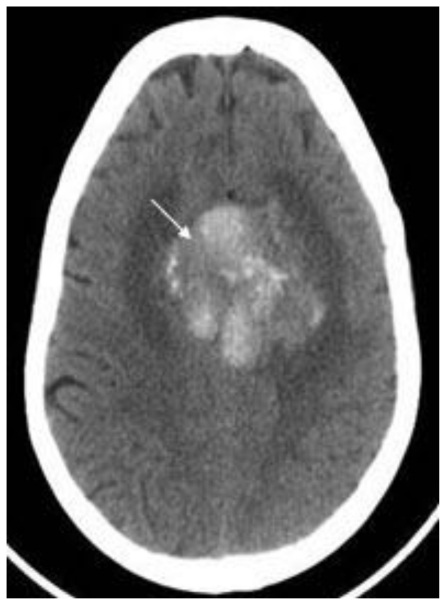
Figure 2.
68 year old female with stage 4 colon adenocarcinoma metastatic to the lungs. Axial noncontrast CT of the brain in demonstrates a 5.2 cm × 4.2 cm × 5.2 cm heterogeneously hyperdense mass in midline of the vertex with calcifications (arrow) suspected to represent a meningioma. August 2008. GE Lightspeed 8-slice CT scanner.

