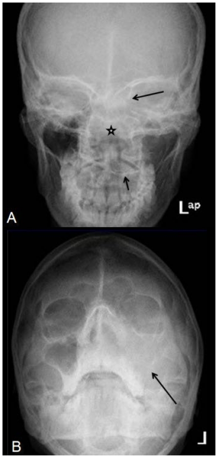
Figure 1.
25-year-old male patient with cemento-ossifying fibroma of the paranasal sinuses presenting with left orbital and periorbital redness and swelling. A: AP Skull radiograph (upper image) shows expanded nasal cavity (asterix) with depressed left half of hard palate (short black arrow) and ill-defined left medial orbital wall (black arrow). The ethmoid sinus trabeculae are not delineated. B: Occipitomental view (lower image) shows soft tissue opacity occupying the left nasal cavity and left maxillary sinus region with nonvisualized maxillary sinus walls (long black arrow).

