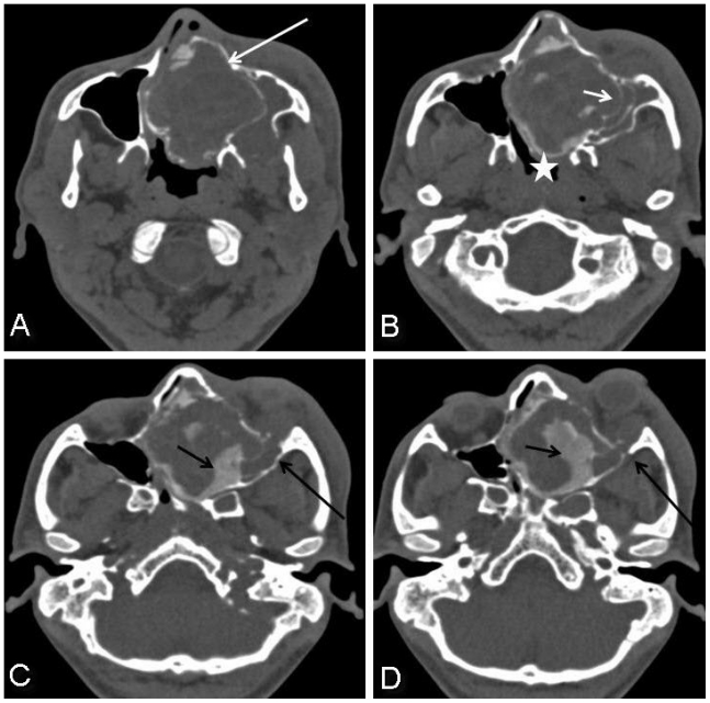
Figure 2.
25-year-old male with cemento-ossifying fibroma of paranasal sinus presenting acutely as orbital cellulitis. Axial noncontrast CT paranasal sinus images in bone window reveal a large, expansile, well-circumscribed, corticated left sinonasal mass involving the left ethmoid sinus, nasal cavity and left maxillary sinus (2a, white arrow). The mass is remodelling the left maxillary sinus medial wall (2b, small arrow) and bulging into the left side of nasopharynx (2b, asterix). There are plaque-like areas of high density (180 – 220 HU) within the lesion suggestive of calcification or ossification (2c, small black arrow and 2d). The lesion extends into the left orbit via inferior orbital fissure causing left orbital proptosis (2c, long black arrow and 2d).

