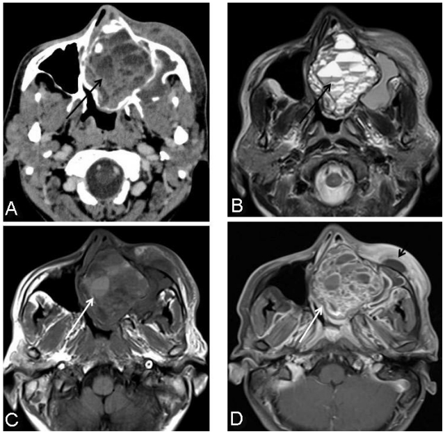
Figure 4.
25-year-old male with cemento-ossifying fibroma of paranasal sinus presenting acutely as orbital cellulitis. Axial post contrast CT paranasal sinus reveals multiple pockets of nonenhancing areas of hypodensity with likely fluid-fluid levels (4a, black arrow). Axial T2 weighted image at the same level confirms multiple fluid-fluid levels (4b, black arrow). Few of these areas are showing T1 hyperintense signal also representing thick secretions or blood-blood levels (4c, white arrow). Post contrast axial T1 fat suppressed image shows irregular rim and solid areas of enhancement (4d, white arrow) with associated premaxillary soft tissue thickening and collection (4d, short black arrow).

