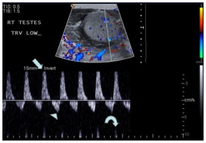
Figure 7.
50-year-old with right sided epididymo-orchitis. Spectral waveform on color Doppler imaging 2 days after admission demonstrates reversal of diastolic blood flow. The peak systolic flow is elevated (arrow), and the peak diastolic flow is reversed (arrowhead). Incidentally noted is aliasing artifact (curved arrow). In the setting of infection, this type of waveform is concerning for impending infarction. Also demonstrated is the previously described complex right hydrocele, representing a pyocele.

