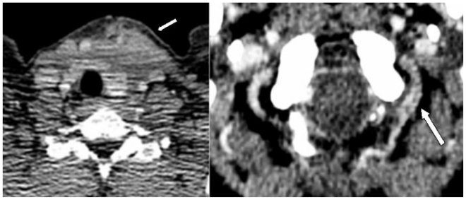
Figure 2.
40 year old women with vertebral arterio-venous fistula secondary to penetrating cervical trauma. Axial Contrast- Enhanced Computed Tomography of the neck demonstrating left anterior neck haematoma (arrow, left) and enhancement of the left vertebral artery at the atlanto-occipital level (arrow, right).

