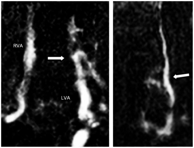
Figure 4.
40 year old women with vertebral arterio-venous fistula secondary to penetrating cervical trauma. Coronal 2D time of flight MRA demonstrating (left) early occlusion of the left vertebral artery (white arrow) with fistulous connection. A further coronal image (right) shows fistulous connection and arterial flow in a left vertebral vein (white arrow). RVA = Right vertebral artery, LVA = Left vertebral artery.

