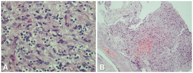
Figure 6.
68 yrs old female with glioblastoma of the optic pathways. A - Photomicrograph showing anaplastic tumor cells in a fibrillary background - tumor cells show abundant cytoplasmic clearing - somewhat reminiscent of ‘fried-egg’ appearance. However, cellular pleomorphism was pronounced (H &E, 400X). B - Photomicrograph showing focal necrosis and hemorrhage within the tumor tissue (H & E, 100X).

