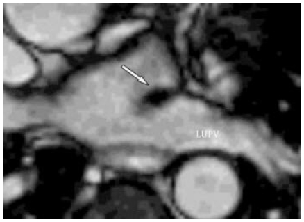
Figure 2.
In this 50 year old female with a prominent coumadin ridge, a customised axial view using a multi-phase balanced SSFP cine sequence (TE 1.7ms, TR 3.5ms, flip angle 60 degrees, SENSE factor 2, matrix 192 × 192, field of view 320–460mm, slice thickness 6mm, 24 phases per cardiac cycle, 1 slice per breath-hold) demonstrates the close proximity of the structure to the left upper pulmonary vein (LUPV).

