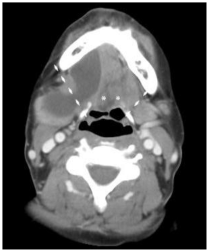
Figure 3.
Initial contrast enhanced axial Computed Tomography - 100 cc of Omnipaque-300 (KVp 120, mA 20 * 3572ms). 44-year-old female with plunging ranula. A more inferior level, at the level of the hyoid bone. The geniohyoids (labeled with “*”) can be appreciated inserting on the hyoid bone. The dashed lines show the posterolateral free-margins of the mylohyoid muscle. The lesion measures 6.7 × 2.2 × 4.5 cm and is of water-density. It is non-enhancing, homogenous, smoothly-marginated, and without internal septations. The posterior aspect of the lesion can be seen extending posteriorly over the approximate location of the free edge of the mylohyoid bone.

