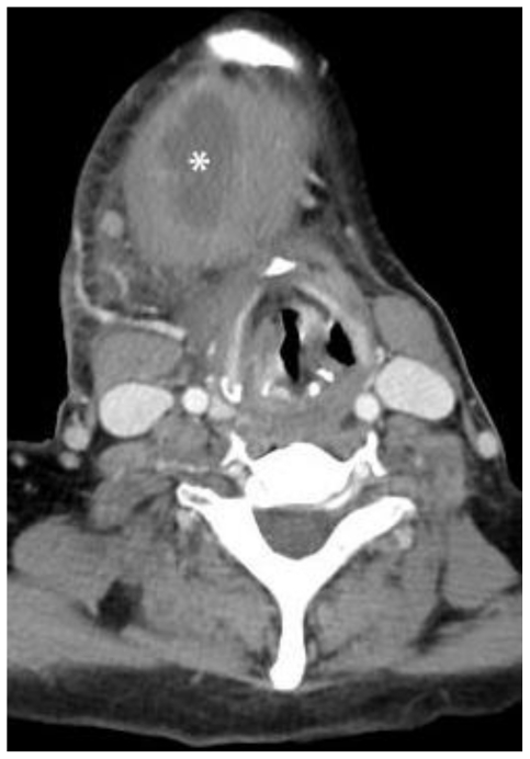
Figure 7.
Follow-up contrast enhanced axial Computed Tomography performed 2 months later - 80 cc of Omnipaque-300 (KVp 120, mA 20 * 3575 ms). 44-year-old female with infected plunging ranula. The image demonstrates the inferior aspect of the lesion. The mass (labeled with *) has unchanged anatomic relationships, but has slightly enlarged and now measures 6.9 × 3.7 × 4.0 cm. It remains homogenous, smoothly-marginated, and without internal septations. Thick rim-enhancement is now seen, consistent with secondary infection. Mass effect with tracheal deviation and compression is worse and there is new right submandibular and cervical reactive lymphadenopathy. The mass was surgically excised and pathologically proven to be a plunging ranula.

