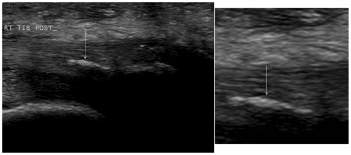
Figure 3.
A 42 year old female was diagnosed with calcific tendinosis of the posterior tibialis tendon. Ultrasound scan of the tibialis posterior tendon carried out using an 8–12 MHz linear array ultrasound probe in the longitudinal plane (slightly different angle than seen on figure 2) showed calcific tendinosis within the tendon distally, close to its insertion into the navicular bone. The rest of the tibialis posterior tendon was intact and of normal echo texture. White arrow indicates location of pathology.

