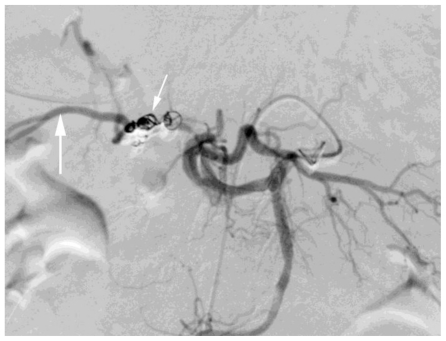
Figure 5.
78 year old female with a cystic artery pseudoaneurysm. Digital subtraction angiogram performed via celiac axis injection. Post embolisation image illustrating successful treatment of the cystic artery pseudoaneurysm with microcoils (small white arrow). Note the celiac axis (black arrow) and collateral filling of the right hepatic artery (large white arrow).

