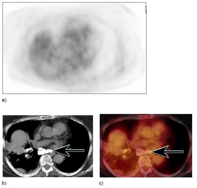
Figure 4.
Large calcified lymph node without AC artifact in an 85-year-old female with questionable soft tissue abnormality in right hilum. Scan was performed 60 minutes after injection of 12–16 mCi of FDG tracer, with the following parameters: 120 kVp, 300mAs, 5 mm slice thickness, and CT based attenuation correction algorithm using two iterations and 8 subsets. Axial AC PET shows no FDG activity in the area of large subcarinal calcified lymph nodes, as seen on axial CT and AC axial fused PET/CT images (b,c) (HU: min 520, max 1371, mean 809).

