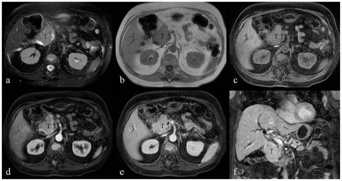
Figure 3.
66-year-old male patient presented with severe nausea and vomiting in the setting of a two-week history of worsening fatigue, pruritus, and jaundice. a: Transverse noncontrast, T2-weighted, fat-suppressed image (TR/TE = 2600 msec./160 msec.) demonstrates the duodenal tumor showing homogenous high signal intensity. b: Transverse noncontrast, T1-weighted, 2D fat- suppressed spoiled gradient-echo sequence (TR/TE = 177 msec./4.3 msec.; flip angle = 80 degrees) demonstrates the duodenal tumor showing homogenous signal intensity isointense to the paraspinal muscles. c–f: Transverse, T1-weighted, fast-suppressed, 3D spoiled gradient echo (LAVA) sequences (TR/TE = 4.216 msec./2.02 msec. flip angle = 12 degrees), before and after intravenous contrast (20 ml gadobenate dimeglumine, Multihance). Duodenal tumor demonstrates mild enhancement during the hepatic arterial-dominant phase (d), which becomes homogenous and more intense after one minute (e), and two-minute delay (f). T: duodeal tumor.

