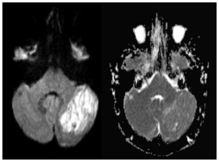
Figure 2.
41 yo female with history of multiple sclerosis presented with horizontal diplopia. Axial diffusion weighted imaging (left image, B-1000)(1.5 Tesla Siemens Espree, repetition time (TR) = 4500 ms; echo time (TE) = 112 ms) demonstrates a 3 × 4 cm large, well defined area of predominantly corduroy appearing, increased signal in the left cerebellar hemisphere with sparing of the cerebellar peduncle, causing slight mass effect upon the left aspect of the 4th ventricle. Corresponding apparent diffusion coefficient (ADC) map (right image) shows increased diffusion values proving lack of diffusion restriction. This excludes acute ischemia and is typically found in LDD. Medulloblastoma that may present with diffusion restriction becomes a less likely differential diagnosis.

