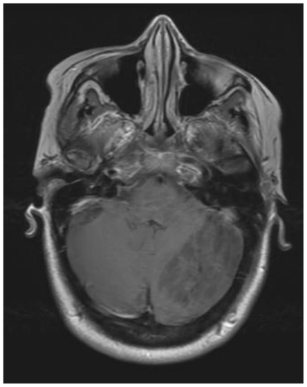
Figure 3.
41 yo female with history of multiple sclerosis presented with horizontal diplopia. This axial T1-weighted image (General Electric Signa 3.0Tesla; TR = 2607ms, TE = 9.94 ms, inversion time (TI) 1100ms, number of excitations (NEX), 2; flip angle FA = 90) demonstrates alternating isointense and hypointense signal of the tumor within the left lobe of the cerebellum. This is the characteristic striated corduroy pattern of a tumor in Lhermitte-Duclos disease.

