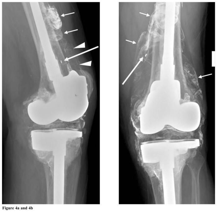
Figure 4a.
and 4b. These “off lateral (a = left)” and AP (b = right) radiographs of the left knee were obtained about 18 months after the Fig. 3 images. There are new lobulated and somewhat bubbly densities around the distal femur (short white arrows in Figs. 4a and b) similar to the “bubble sign” that has been described after total hip arthroplasty. Linear densities are also seen in the suprapatellar region on the lateral view (arrowheads in Fig. 4a), similar to the “metal-line sign” of metal-induced synovitis. Both the “bubble sign” and “metal-line sign” represent metallic debris outlining the joint space, effectively demonstrating the joint cavity. Also, note that the small metallic density associated with the patellar polyethylene marker has dissociated superomedially (long arrow in Figs. 4a and 4b), compared to Fig. 3. This is an uncommon but important finding of hardware failure.

