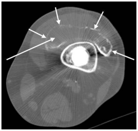
Figure 5.
74 year-old man after left total knee arthroplasty revision with metallosis and metal-induced synovitis. Noncontrast axial CT of the left lower extremity at the level of the distal femur (GE Lightspeed 16 CT scanner; kVp 120; mA 310; 2.5 mm slice thickness) demonstrates linear radiodensities outlining the suprapatellar joint space (short arrows). The joint cavity is distended and outlined by increased density. This finding corresponds with the “metalline sign” seen on plain radiographs (see Fig. 4a), consistent with metal-induced synovitis. There is relatively low attenuation within the joint space. The Hounsfield unit measurement in this region is limited by streak artifact from the adjacent hardware, but the area that the long arrow points to in this image measured 100 Hounsfield units.

