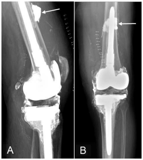
Figure 8.
74 year-old man with metallosis. Lateral (Fig. 8a) and AP (Fig. 8b) radiographs of the left knee show interval removal of the metal-backed patellar component and dissociated liner (reference Figs. 4a-d). A subtotal synovectomy and metallosis debridement were also performed with removal of the bubbly and linear radiodensities that were seen around the distal femur (reference Figs. 4a-d). Dense residual debris and/or calcification is noted in the highest portion of the suprapatellar joint cavity (arrows), and a joint effusion and skin staples are present.

